Product Details
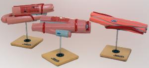
Ward’s® Muscle Types Model Set
The three-model set displays the differences between cardiac, skeletal, and smooth muscle structures.
Description
- Durable Plastic Resin Construction
- Each Model Has Individual Reference Key
- Models Mounted on Individual Bases
The cardiac muscle cell, enlarged 1500 times, is dissected to display syncytial fiber arrangement and Purkinje cells; the skeletal muscle, enlarged 1000x, shows the union with both and tendon, and sarcolemma; and the smooth muscle, enlarged 2500x, exhibits the capillary and motor end plate of the autonomic nerve. All models are cross sectioned to display myofibrils.

