Microscopy
Showing all 15 results
-
Biology
Human blood, microscope slide
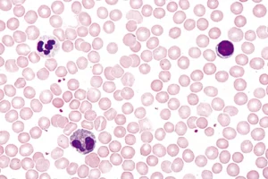
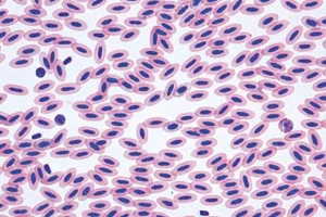
The microslide shows a blood smear of human blood that has been fixed and stained so that the different blood cells can be clearly distinguished. The red blood cells appear red, while the white blood cells are seen in purple-blue shades with different sizes and nuclear types. The slide provides a clear insight into the composition of the blood and makes it possible to work with both the structure and function of the individual cell types.
SKU: FR-079145 -
-
-
Biology
Microscope slide set, botany, 25 different
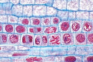
Affordable botanical basic set with 25 different micro-slides from the world of botany. The slides are mounted on glass slides and cover a wide range of plant tissues and microscopic structures that you and your students can examine under a microscope.
SKU: n/a -
Biology
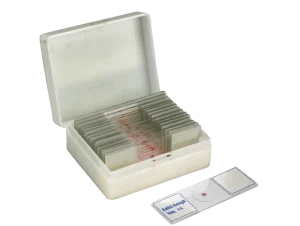
Microscope slide set, histology, 25 specimens
This affordable basic histology set contains 25 microscopic slides from a variety of mammals and vertebrates such as humans, dogs, rabbits, frogs and mice. The histology set is designed for educational use and allows students to study the structure of different tissue types such as connective tissue, muscle tissue, nervous tissue and epithelium.
SKU: FR-078854 -
Biology
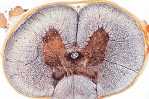
Microscope slide spinal cord of cat, t.s.
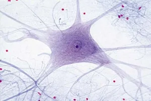
Micro-preparation of spinal cord from cat, cross-section. The preparation is made by Lieder in high quality. Each preparation is unique, and natural variations may occur, which means that the color and shape may differ from the images shown. The preparation is ideal for use in teaching to give students a concrete understanding of the anatomical appearance of the spinal cord.
SKU: FR-079225 -
Biology
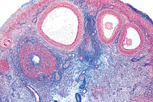
Ovary, cross-section, microscope slide
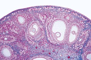
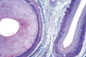
Single slide of an ovary showing the structures and cells that play a role in a woman’s follicle maturation and ovulation. The appearance of a micro-slide of the ovary is very similar to the illustrations you see in a biology book, so it does not require any medical training to find the structures and cells you have read about.
SKU: FR-079200 -
-
Biology
Testicle, cross-section, microscope slide
This microslide shows a cross-section of testicular tissue, showing the cells and structures involved in sperm formation. The slide is stained with red and blue contrast dyes, making it easier to distinguish the different cell types and tissue layers. The nuclei of the cells appear as darker structures, while the cytoplasm and cell membranes form the surrounding shapes. The slide is designed to closely resemble illustrations in biology textbooks, making it easy to recognize the most important details, even without a medical background.
SKU: FR-079205