
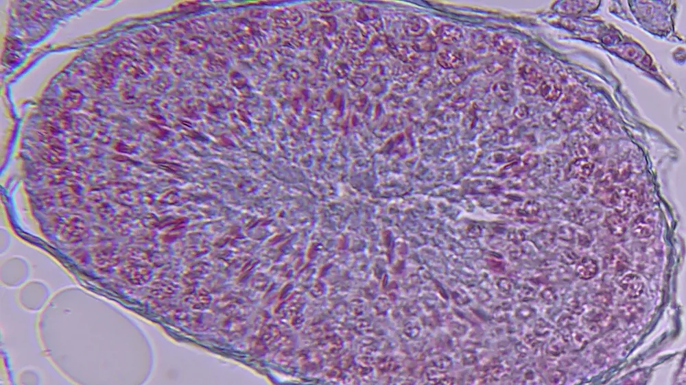
Testicle, cross-section, microscope slide
This microslide shows a cross-section of testicular tissue, showing the cells and structures involved in sperm formation. The slide is stained with red and blue contrast dyes, making it easier to distinguish the different cell types and tissue layers. The nuclei of the cells appear as darker structures, while the cytoplasm and cell membranes form the surrounding shapes. The slide is designed to closely resemble illustrations in biology textbooks, making it easy to recognize the most important details, even without a medical background.
In biology lessons, the microslide allows students to examine and identify the central elements of the testicular structure. With a microscope that can magnify around 60x, students can clearly see cross-sections of the seminiferous tubules, basement membranes, lumens, and various cell types such as sperm cells, germ cells, Leydig cells, Sertoli cells, and fibroblasts.
The specimen supports work on topics such as human anatomy, reproduction, cell differentiation and hormone production. Students can use it to connect theoretical knowledge from textbooks with practical observations under the microscope. This strengthens their understanding of how different cell types work together in the process of sperm production and how tissues are organized within an organ.