Product Details
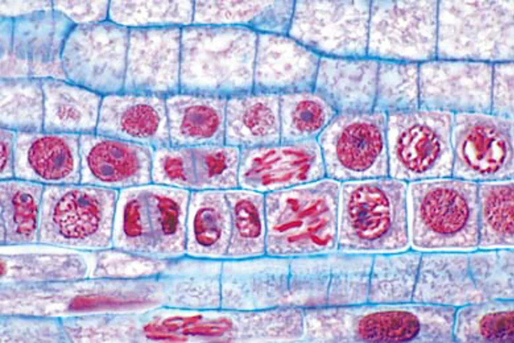
Slide, DNA, cell division
This microslide shows mitotic cell divisions in the root tip of onion and is prepared to illustrate stages of mitosis.
Description
The slide is fixed on a slide and stained with a quadruple staining method using safranin, methyl violet, fast green and orange G. This staining provides high contrast and clear visualization of the cell nucleus, nuclear membrane, nuclear bodies and chromosomes, so that the individual phases of cell division appear clearly under the microscope.
Use of the product
In biology lessons, the microslide can be used to study and identify the different stages of mitosis – prophase, metaphase, anaphase and telophase. Students are given the opportunity to connect theory about DNA and cell division to concrete observations through microscope work. The slide also supports work with basic laboratory techniques and gives students experience in interpreting biological structures on microscopic slides.
In professional contexts, preparations like this are used in laboratories and educational institutions for teaching and training in cytology. It can also be used in courses within biotechnology and health education, where understanding cell division is central to both research and diagnostic work.