Product Details
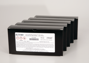
Animal cells, 12 microslides
Series of 12 selected preparations of various animal cell tissues. Supplied in a preparation box.
Description
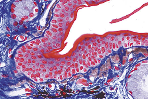
Squamous epithelium Mucous membrane (human). Isolated cells with stained cell nuclei and cytoplasm from the oral cavity
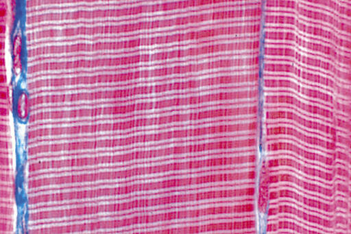
Striated muscle tissue – Structure of tissue in striated, myofibrils and muscle tissue cell nuclei. Longitudinal section
Bone tissue – Compact with hyaline cartilage tissue. 2 sections on the same slide for comparison. Cross section
Nerve fibers – Isolated, fixed and stained to show myelin sheaths and constrictions of Ranvier
Liver tissue – Salamander. Cross section
Kidney Tissue – Mouse. Stained to show storage in the epithelial cells. Cross section
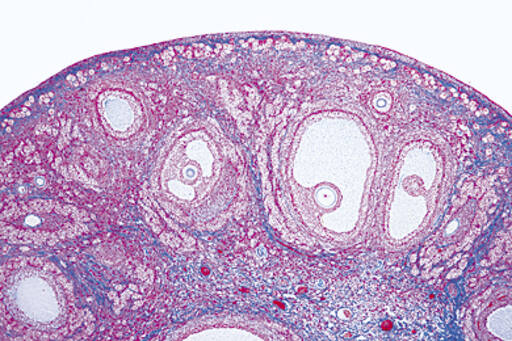
Ovary – Cat. primary, secondary and graafian follicles. Cross section
Testicles – Seeds. Spermatogenesis: spermatogonia, spermatocytes, spermatids and spermatozoa. Cross section
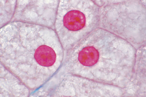
Mitosis – Salamander larvae. Skin and other organs show mitosis in different stages. Cross section
Uterus – Horse roundworm. Colored to highlight details chromosomes and filaments at meiosis. Cross section
Giant chromosomes – Dancing mosquito larvae. Salivary gland tissue is stained to highlight DNA and shows giant chromosomes and chromomeres.
Cleavage – Sea urchin. Unfertilized and fertilized egg show different cleavage stages.

